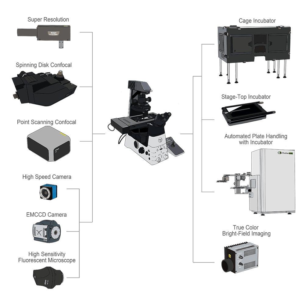
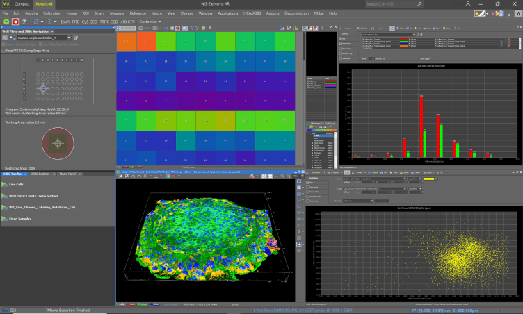
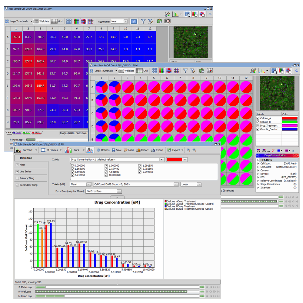
A study of living cells using microscopy to obtain a better understanding of biological function through the study of cellular dynamics.
With Nikon microscope and software NIS-Elements, our high content imaging streamlines high-speed imaging and simple operations.
Automated well-plate acquisition, data review, analysis, and management for multiple well-plate experiments.
Real-time viewing of data acquisition and analysis progress for instant inspection.Multiple analysis assays can be run simultaneously during the imaging phase or run post-acquisition on offline stations.
Compatible with various microscopy, including Confocal, Fluorescent, Phase Contrast, or Differential Interference Contrast Microscopy, cameras, and incubators.
Easy define acquisition parameters, including well-plate configuration, plate handling, autofocusing, filter switching, and detectors
With the Nikon Perfect Focus System (PFS), focus drift is corrected automatically caused by
temperature changes and mechanical vibrations, which is due to a variety of factors including the addition of reagents to the sample and multi-position imaging.
The PFS enables faithful visualization of the nanoscopic and microscopic world during long time-lapse experiments.
Fast and efficient clarity in images which is normally corrupted by blur due to out-offocus light through a pre-trained algorithm. Clarify AI can be applied on any widefield 2D or 3D data set, detector, or magnification.
By tracing features of interest and training these compared to the underlying image of Segment AI, segmentation can be applied to similar images, accurately set up the cell region through various criteria, which were previously only identifiable by painstaking manual tracing approaches.
Enhance.ai enables cell counting or segmentation without stained or harmful light excitation. The network learns the patterns in Bright-field images and predicts the
details in the fluorescent channel.
By Convert AI, segmentation is applied automatically on label-free approaches, or imaging without harmful near UV excitation if the AI is trained to predict the specimen pattern in a particular channel. Throughput of acquisition as well as specimens viability both increase as a result.
Denoise.ai can recognize and remove the shot noise component of confocal images, increasing clarity and allowing for shorter exposure times.
*Photos courtesy of Nikon official website
Our High Content Imaging System applies to a variety of researches. For instance, Live Cell Imaging, Large Tissue or Organ Investigation, Drug Screening, etc.
Defective apoptosis is implicated in various diseases such as cancer, neurodegenerative diseases, and autoimmune diseases. Drug efficacy tests using apoptosis as an index are widely used in drug screening. Pharmacological tests require consideration of various conditions such as drug concentration and treatment time, but the efficiency of experiments can be improved with time-lapse imaging since it can obtain multiple time point data using only one well-plate. In addition, since live-cell assays can analyze the same cell population over time, results can be obtained with a minimum of variability.
A long-term live-cell apoptosis assay with low phototoxicity is used in this experiment. Low phototoxicity was achieved by generating a phase distribution image (Volume Contrast image) that reflects the cell structure from multiple brightfield images captured in different focal planes, and by measuring the total number of cells over time in the field of view, enabling a long-term apoptosis assay.
Analysis results showing that apoptosis was induced in a concentration-dependent manner for staurosporine (STS) VC images were overlaid with a cell classification mask.
Blue: Non-apoptotic live cells
Green: Apoptotic cells
Red: Dead cells
Upper row: STS OuM
Middle row: STS 0.5 UM
Lower row: STS 10 UM
From the left: Oh, 12h, 24h, 36h, 48h after drug was added
Scale bar: 200 um
can be quantitatively evaluated by the ratio of the number of apoptotic cells to the total number of cells. In short-term live-cell assays, nuclear stain reagents such as Hoechst are used for measuring the total number of cells, but in long-term assays, cell proliferation is suppressed by dye toxicity and fluorescence excitation phototoxicity, and this causes a decrease in reproducibility in the quantitative analysis of drug efficacy.
With Nikon microscope and software NIS-Elements, our high content imaging streamlines high-speed imaging and simple operations. Nikon software provides a dedicated interface for high content acquisition and analysis routines.
Without the need to adjust the focusing method, wavelengths, filters, and camera settings, it is easier to process alongside your research quickly and effortlessly.
*Photos courtesy of Nikon official website
Designed with Mobirise
Free Offline Web Page Builder