See Brighter, See Faster, See More
High-sectioning and High-speed in 25mm Large Field of View makes X-Light V3 capable for the modern fluorescence microscopy applications.
The first confocal unit which allows dual camera imaging at the full field of view of 25 mm on both cameras.
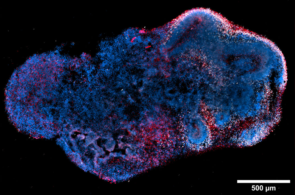
Large image of a day 50 human
cerebral organoid
Micro-lenses make the illumination field highly homogeneous on large fields
of view (up to 25mm). With large FOV and high-speed imaging
(15,000 rpm), quantitative imaging on large samples becomes more
simple.
Flexible switching between different illumination sizes to fit your application.
Achieving higher power density for higher speed imaging (1KHz) is
possible by changing to a smaller illumination area.

- Microscope (Nikon Motorized Inverted Microscope Ti2-E, with 25mm FOV)
- Illuminators (Lumencor)
- Detectors (Andor)
- Software (Nikon NIS-Elements)

Brain organoid derived from human iPSC line (MAP2-AF488, labels microtubules in neuronal dendrites, TBR1-AF750 labels nuclear
transcription factor, and DAPI stains DNA
Dual camera imaging at the 25mm full field of view on both cameras
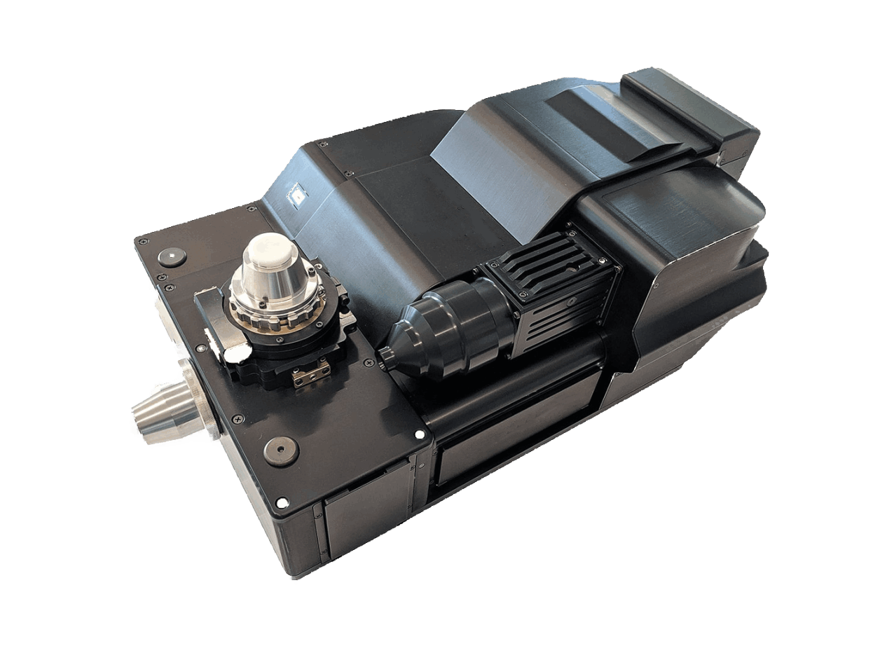
Makes the Spinning disk fully isolated and protected from environmental dust and prevents undesired artifacts related to the presence of small dust particles on the Spinning disk surface
*Photos and Videos courtesy of CrestOptics official website

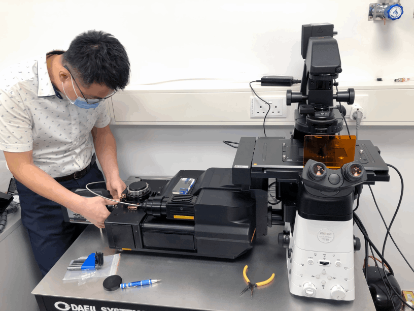
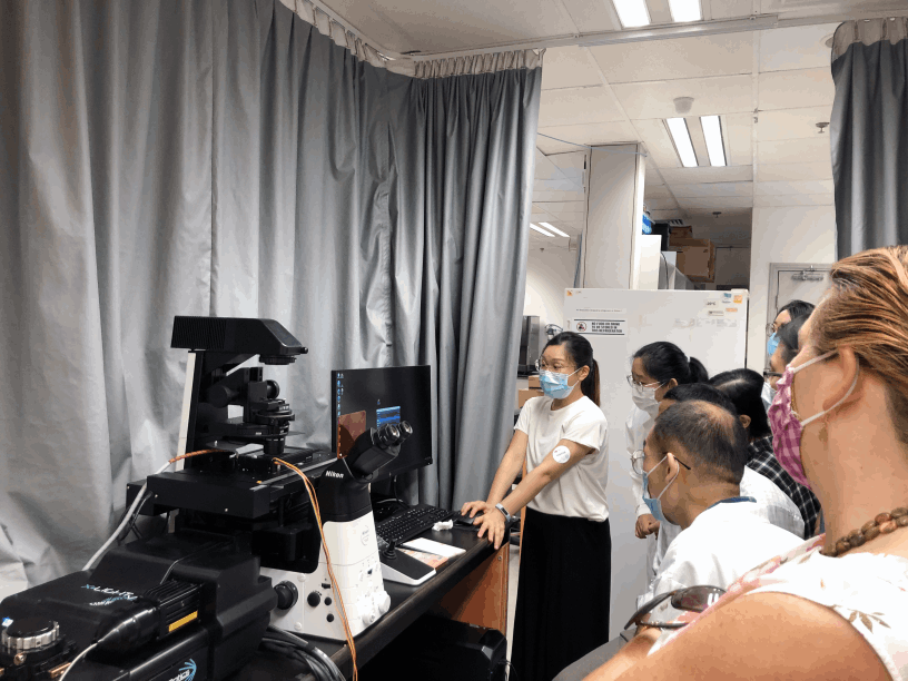
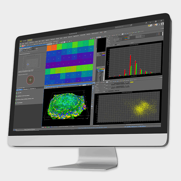
With Nikon microscope and software NIS-Elements, our high content imaging streamlines high-speed imaging and simple operations. Nikon software provides a dedicated interface for high content acquisition and analysis routines.
Without the need to adjust the focusing method, wavelengths, filters, and camera settings, it is easier to process alongside your research quickly and effortlessly.


Designed with Mobirise - Go now